Superficial Siderosis
Note extensive hemosiderine deposits superficially around brainstem, cerebellum - seen on first T2 images as well as deposits round basal ganglia and on the brain surface on the GRE (Gradient Echo) images.
Note extensive superficial hemosiderine deposits at cerebellum surface and brainstem on above GRE images.
Above coronal, sagittal, axial T1 and coronal FLAIR sequences show extensive cerebellar atrophy - that is also characteristic to Superficial Siderosis.
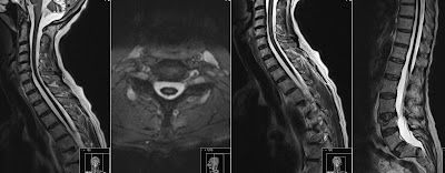
Note also extensive Superficial Siderosis of the spinal cord as well as it's atrophy.
Superficial Hemosiderosis (Superficial Siderosis) of the central nervous system results from chronic iron deposition in neuronal tissues associated with cerebrospinal fluid. Residues of blood are penetrating the pia mater and deposit in the superficial layers of the cerebral cortex. Superficial cortical hemosiderosis is defined as linear residues of blood in the superficial layers of the cerebral cortex. It has been shown in animal models with experimental siderosis that repeated bleeding in the subarachnoid space leads to deposition of hemosiderin in the subpial layer of the brain. When observing low signal deposits of hemosiderine it is important to note if it is located in the subarachnoidal space or superficially at the surface of the central nervous system. Superficial Siderosis is an effect and not a cause. One of the proposed causes is Cerebral Amyloid Angiopathy (CAA).
See also interesting article from AJNR:
Subarachnoid Hemosiderosis and Superficial Cortical Hemosiderosis in Cerebral Amyloid Angiopathy - J. Linn


Post a Comment for "Superficial Siderosis"